Cardiac MRI may have Distinguished Commotio Cordis from Vaccine Myocarditis


One of the best pieces of news for me has been young Daimler Hamlin’s announcement that he is fit to resume playing American football. The young man had collapsed on the field not too long ago after receiving a chest blow from an opponent. There are multiple videos confirming the blow to the chest and Hamlin collapsing soon after. Luckily for him, there were paramedics around who defibrillated him. There is news that he needed more than a couple of DC shocks. He was transferred to a hospital for the necessary observation and management. We have no access to the investigations done, and rightly so.


Many cardiologists suggested that this was a clear-cut case of vaccine-induced myocarditis-induced sudden death syndrome because of the vaccine mandate in his football league. There have been multiple reports of young people, especially men, collapsing on the playing field, with some sadly dying.


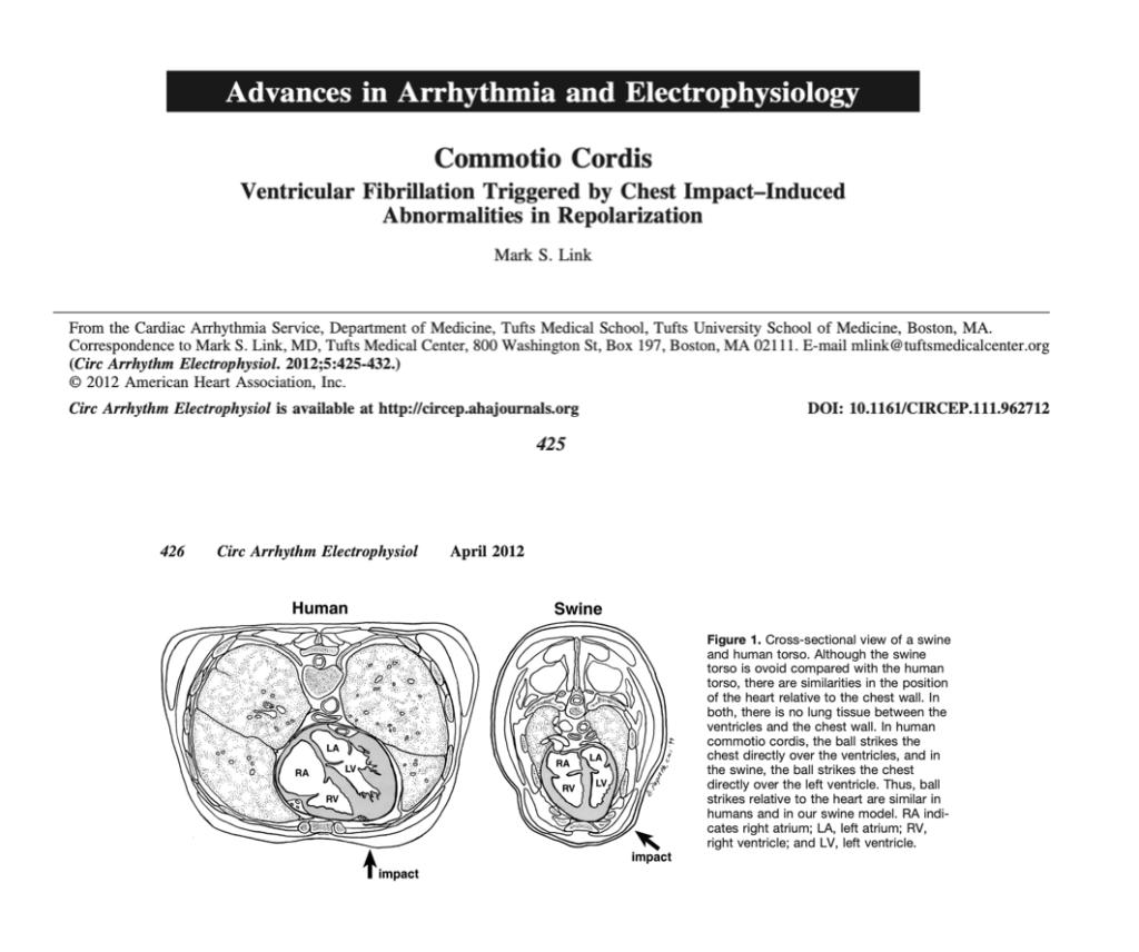
In the case of Hamlin, the presentation is quite different; there is a definite blow to the chest, following which Hamlin collapses after a few seconds. The sequence of events confirms Commotio Cordis. The unfortunate blow was timed to perfection, some milliseconds before the peak of the T wave, and with sufficient force to trigger a polymorphic ventricular tachycardia that soon degenerated into ventricular fibrillation.


On the sporting field, commotion cordis is seen more commonly in men than in women; the explanation given is that men may have a lower threshold or reserve during the depolarization phase. However, outside of the sporting field, more women suffer than men. The situations leading to Commotio Covid could be a motor vehicle accident, a brawl, or some accident in which the chest is struck by some object.


Experiments in animals have revealed that in a 20-kg swine, a pressure impact of 300 mmHg to 500 mmHg at a speed of 30 to 50 mph would be enough to trigger an unstable heartbeat or a ventricular tachyarrhythmia. The timing of the impact is crucial; it should be during the vulnerable period just before the T wave of the ECG peaks. This could be 30 to 40 milliseconds earlier.


There is little tissue between the human heart and the breastbone or the ribs. The bones in the young are still in development, hence Commotio Cordis is most often reported in teenagers aged 16–17 years. An underdeveloped sternum makes the heart more susceptible to a lethal physical impact.


Any sport entailing physical contact or the hurling of a hard ball can lead to an incident. Baseball is a good example, where a firm and hard ball is thrown towards the batter. There can always be a chance of the ball being missed by the baseball bat and striking the batter’s chest. The game of cricket is different. The cricket ball has to hit the pitch first before bouncing off towards the stumps. The chances of the cricket ball hitting the chest of the batter are therefore considerably diminished. The cricket ball after pitching usually stays knee high, but a bowler could deliberately bowl a bouncer aiming at the head or chest of the batter.


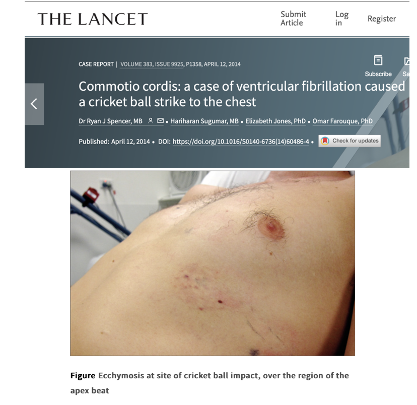
Some years ago, The Lancet published a case report of a 25-year-old engineer who was struck by a cricket ball on his chest; he collapsed about 30 seconds later. Luckily, CPR was initiated, and defibrillation was performed as late as 9 minutes after the collapse. The batter regained consciousness, and there were no changes in his ECG, 2-D echocardiogram, or even MRI. The engineer resumed playing cricket after three months.


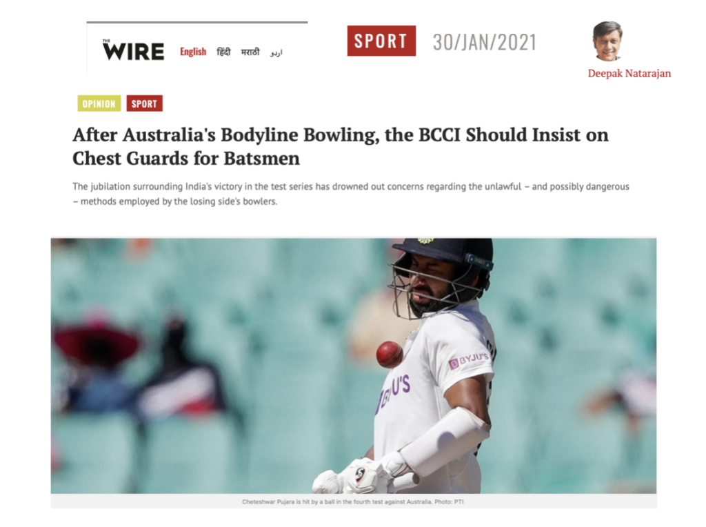
I had written an article for the Wire on the need for the Indian cricket team to wear a chest protector. The article was written during the last Border Gavaskar test series between India and Australia, which took place in Australia. India won the series despite the Australian fast bowlers deliberately aiming for the bodies of the Indian batters. There is no proof that a chest protector will make one foolproof against Commotio cordis, but it just might mitigate the impact.


I would bet that Hamlin suffered from commotio cordis despite being fully vaccinated. For all practical purposes, the vaccine must have been of the mRNA type. For all you know, he could even have underlying myocarditis or heart inflammation because of the vaccine. There is yet another possibility: that he suffered a viral myocarditis some years ago and carries a scar in his heart due to that inflammation. There are case reports of people suffering from commotio cordis over an underlying old myocarditis scar. The scar tissue could serve as a substrate or sensitise the trigger for a dangerous ventricular arrhythmia. This could also occur after an mRNA vaccine-induced myocarditis.


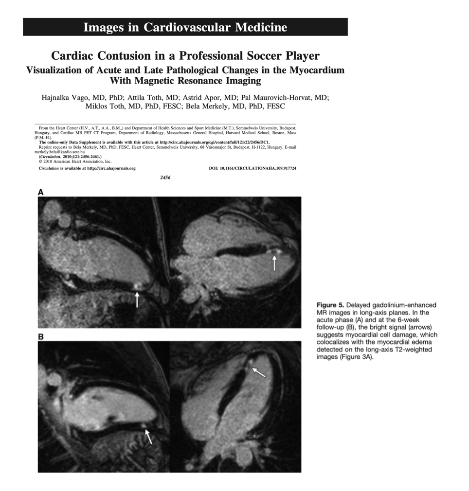
So what do we conclude? The answer is difficult from the outside. The working diagnosis will be commotio cordis because the collapse occurred very soon after being struck in the chest. A cardiac MRI would be of great help in confirming what actually took place. The cardiac MRI may not be definitive if done after 3 months, but if it was done when Hamlin was hospitalised, it could display edoema and haemorrhage in the left ventricle wall. Cardiac MRI presents a distinct picture depending on the pathology. Damage due to an ischemic insult or heart attack always extends from the subendocardium, which is the layer of the ventricle wall lining the cavity. Viral myocarditis extends from outside to inside, from the subepicardium to the midmyocardium. Trauma to the heart could also be subepicardial, but it will be accompanied by a little bleed in the ventricle wall. Both the left ventricle and right ventricle could be involved, with the former being the victim more often.


Remember, Commotio Cordis can occur with minimal trauma to the heart; hence, there may not be too much damage seen, such as avulsion of heart valves. Cardiac MRI, if done soon after the index event, could provide evidence of slight trauma and also rule out acute myocarditis or previous myocarditis that has left a residual scar. Late myocarditis, fibrosis, or scarring is detected by late gadolinium enhancement.


Enough of technicalities; the point I am raising is that to propose Hamlin is a clear-cut case of vax-induced myocarditis resulting in collapse is stretching it a bit too much. It is agreed that many young athletes have collapsed these past two years since the rollout of vaccines, and that many, if not most, of them are due to vaccine-induced myocarditis. But Hamlin’s may not be a vax-induced collapse. Circumstantial evidence favours Commotio Cordis; a cardiac MRI could have cleared the air. An endomyocardial biopsy soon after a traumatic injury may not have been the wisest course of action, but an MRI would certainly have been helpful. Crucially, cardiac MRI will not pick up every case of myocarditis or myocardial injury, and signs of fibrosis or scar tissue will not persist beyond 6 months in quite a few.


If myocarditis due to an mRNA vaccine or previous virus infection is strongly suspected or confirmed, an implantable cardioverter-defibrillator (ICD) becomes necessary to prevent a future catastrophe.

THE VACCINATION JUGGERNAUT IS RUNNING OUT OF GAS


JAMA never stops surprising me. It keeps putting out articles about SARS Cov 2 that are so funny they make me laugh out loud. The papers published on ivermectin have glaring conflicts of interest that are clear as day to everyone but the editors of this medical journal. The ivermectin papers are designed to convince you that, out of the hospital, ivermectin is no better than a lump of sugar in treating COVID patients. Invariably, the drug is given late after symptom onset, on an empty stomach (it should be taken with food) , or the diagnosis of COVID itself is ambiguous. It is well known that the authors have been provided millions of dollars to do the research, apart from either being funded or employed by a company making a competing medicine.


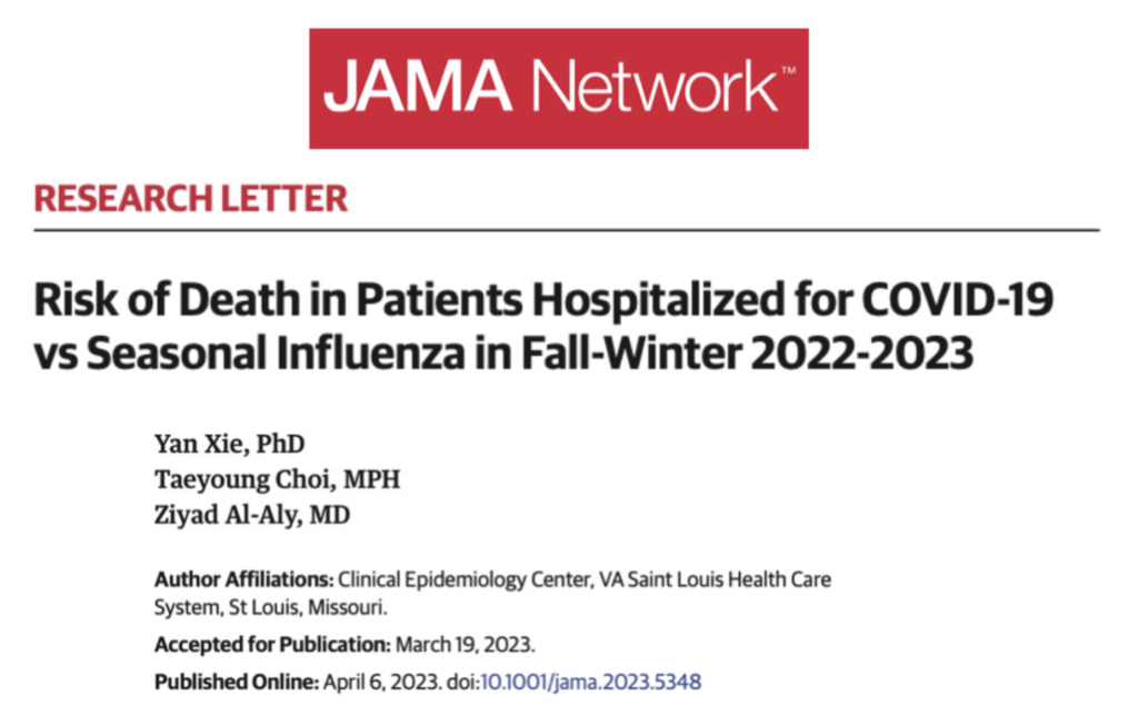
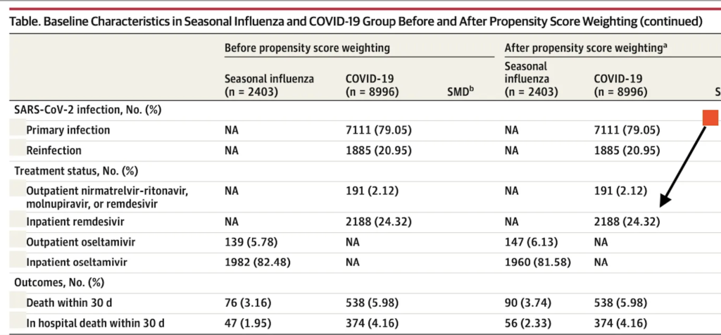
The latest example is a scientific letter that concludes that mortality in hospitalised patients with SARS-CoV-2 is greater than that of those admitted with the flu. This is based on data from the US Department of Veterans Affairs. Elaborate tables are provided with elaborate statistical analysis after propensity score matching of both groups.


But on closer inspection, one realises that more than one fourth of patients received remdesivir. The patients being analysed were admitted at the end of 2022. It is well known that remdesivir killed more than 50% of ebola patients in a study comparing it to other drugs. The paper was published as recently as 2018. Fauci knew that the drug was deadly even as he touted it from within the White House.


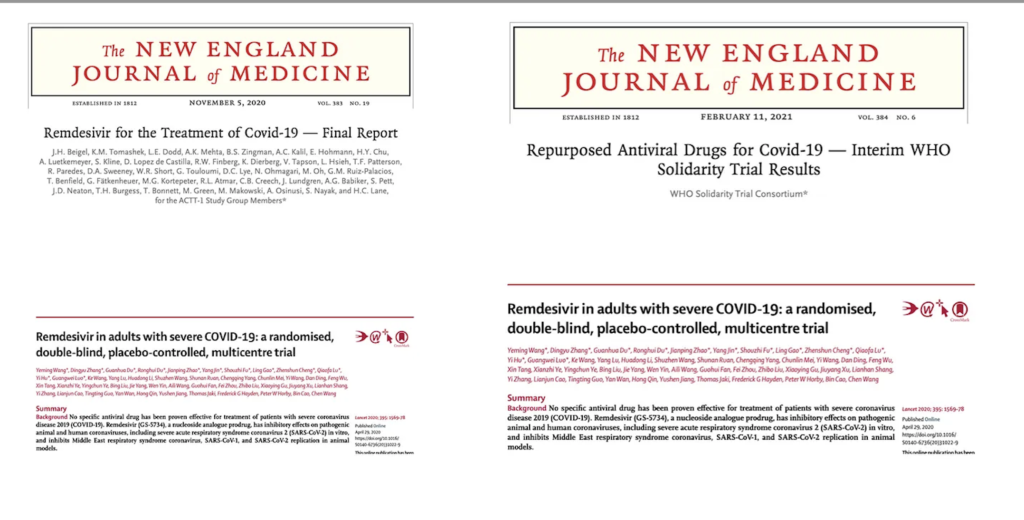
A Chinese study found no evidence that remdisivir helped people in the clinic. A French study came to the same conclusion. An NIAID-funded American study too was unable to document a reduction in mortality; all it could accomplish was a quicker recovery by a few days but no significant reduction in mortality. Even the WHO, to everyone’s surprise, reported their SOLIDARITY trial, which also found no clinical benefit with remdesivir. These trials were published way back in 2020 and 2021.


So to find that remdesivir continues to be used in American hospitals is a mystery.


The point that I am trying to make is that when 24% of people in the COVID group are given toxic medicine compared to none in the flu group, there is bound to be a huge confounder. The playing field is already stacked against the poor COVID patients as compared to the flu cohort in terms of mortality.


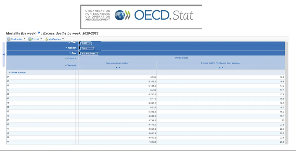
The conclusion drawn by the authors that COVID is more lethal than the flu is, therefore, null and void. It is not a fair comparison. Crucially, there was no difference in death in those below 65 years. We also have data showing that there has been excess mortality in the USA throughout 2022 and during the months this study is evaluating. Excess mortality is more than 25 percent in some weeks, despite more than two-thirds of the population being vaccinated.


In this study itself, two thirds of people are vaccinated, yet they still land up in hospitals. More than 63% of patients admitted with flu had been vaccinated against it, so it’s not something to write home about. Similarly, almost 55% of those admitted for COVID had received a booster, so clearly the COVID booster is not preventing hospital admission.


Deaths in those hospitalised were greater in the unvaccinated versus those boosted, 8.8% versus 5.2%. But again, concentrating only on this data analysis and not considering world-wide data (showing greater death rates in the vaccinated), the administration of remdesivir to almost 25% of patients and the denial of any repurposed drug have to be factored in.


“The risk of death was higher for people who hadn’t been vaccinated than for those who had been vaccinated or boosted.” These results show how important vaccination is for lowering the risk of death from COVID-19.


The above conclusion by the authors is flawed because of the significant use of remdesivir in COVID patients. Also, being in the USA, none of these patients must have received early treatment for COVID in any form. Nearly 6% of people with the flu got oseltamivir, but only 2% of people with COVID got a monoclonal antibody. Worse, more than 80% of hospitalised flu patients were given oseltamivir, even though most people know that it is pretty much useless after 48 hours of the onset of symptoms. Not one patient with COVID received a single repurposed drug.


The authors do concede that the cause of death in these patients is unclear; this has been the bane of almost all studies done during the pandemic. It has been impossible to confirm whether the PCR tests done in the USA (with inflated ct values hovering at 40) were accurate in diagnosing COVID patients, and there has never been clarity on whether SARS-CoV-2 actually caused the death or was simply a bystander.


Importantly, how does one tease out deaths due to SARS-CoV-2 from the boosters or vaccines administered to these patients? Vaccine-induced injury is reported as high as 3 percent, while autopsy series suggest deaths due to vaccines are as high as 14 percent.


So all things considered, this scientific paper published in JAMA is a feeble commercial for COVID vaccine boosters, but the data provided strongly suggests that vaccines along with boosters are failing to protect against hospitalisation or death. But worse is the fact that American hospitals continue administering one of the most toxic drugs on the market, making the populace sitting ducks by not providing early treatment with cheap repurposed medicines.


European Society of Cardiology 0/1-hour algorithm for Diagnosis of Myocardial Infarction is Not Fool Proof.


A patient presenting with chest pain can be a diagnostic dilemma. Especially if there is no or little change in the ECG. The bedrock of diagnosing acute myocardial infarction rests on rise and fall in troponin level. With advent of high sensitivity troponin European and Australian cardiology societies have adopted a 0/1 hour algorithm. High sensitivity troponin is assessed at entry into the emergency and then later after 1 hour; if both values are within normal limits, subsequent myocardial infarction or death can be ruled out with great certainty.


High-sensitivity troponin assays are the preferred biomarker for the detection of myocardial injury. Levels are reported in ng/L. The assay-specific 99th percentile upper reference limit is the threshold for the diagnosis of myocardial injury.


The 0/1 hour high sensitivity troponin algorithm has now been been studied in patients with a history of coronary artery disease presenting to Emergency. Patients with known coronary artery disease compared to those without coronary artery disease  were older , and more likely to be male.


The researchers have reported that in the rule out zone , 30 day death or myocardial infarction occurred in 3.4% of patients with coronary artery disease while it occurred in 1.1% patients without. The negative predictive value (NPV)  of 0/1 hour algorithm did not achieve the 99% or more for the primary endpoint. Moreover addition of the History, Electrocardiogram, Age, Risk Factor, and Troponin (HEART) score did not  significantly improve the NPV.


In  the  1462 patients studied NPV of the 0/1 hour algorithm was 99% for an index myocardial infarction and 98.4% for 30 day cardiac death or myocardial infarction. The NPV’s improved with addition of clinical score to 99.5% and 99.3%. The negative predictive value for 30-day cardiac death or MI was 96.6% (95% CI, 92.8-98.8) among patients with known CAD and 98.9% (95% CI, 97.8-99.6) in patients without known CAD (P = .04).


Among patients with known CAD, the ESC 0/1-hour high sensitivity troponin algorithm was unable to safely exclude 30-day cardiac death or MI. This suggests that clinicians should be cautious if using the algorithm in patients with known CAD. The negative predictive value was significantly higher in patients without a history of CAD but remained less than 99%.


With death or myocardial infarction (MI) within 30 days excluded based on this high-sensitivity cardiac troponin  algorithm, people with a known history of coronary artery disease still experienced this outcome in 3.4% of cases.


“This 3.4% missed 30-day death or MI rate is higher than the 1% miss rate most emergency clinicians are generally willing to accept. Thus, our results suggest that the ESC 0/1-hour hs-cTnT algorithm does not safely rule out ACS [acute coronary syndrome] in patients with known CAD,” the authors wrote in their paper published 2 days ago in JAMA.


Hence for the 0/1 hour algorithm to work the NPV must be greater than 99%, and this has been accepted by almost all researchers in this field. The other practical choice of course is to increase the interval for testing of high sensitivity troponin, which may drain hospital recourses.


An integrated approach for risk stratification — including patient history, electrocardiogram, and serial high sensitivity troponin — as recommended in the 2021 American Heart Association/American College of Cardiology chest pain guidelines would be the most pragmatic approach.


To conclude clinical judgement based on experience should always trump over an algorithm created by a cardiology association. Can a 99% value be better than 98.9%, most probably not.

FINALLY STENTING PERMITTED IN HOSPITALS WITHOUT SURGERY ON-SITE.


Finally US interventional cardiology societies agree that percutaneous coronary intervention can be safely done in hospitals without surgical facilities. I have been advocating this for years, because we have achieved more than a fair bit of experience and  the hardware too has evolved considerably. 


I remember the days performing a coronary stenting procedure was considered a Mount Everest climbing feat without oxygen. I never understood the rationale behind the conservative mindset; maybe it was to retain some sort of monopoly. This was, however, at the cost of a patient.


India has a mammoth population ( probably the maximum on the planet) with sadly a large proportion vulnerable to acute coronary syndrome. The Pandemic has added to the cardiovascular sick pool when it put the entire population under house arrest for months. This was a humongous error.


It is well known that a sedentary life style is as dangerous a risk factor as is smoking, hence lockdowns have most certainly aggravated cardiovascular problems.


But now finally, according to a new expert consensus statement from the Society for Cardiovascular Angiography and Interventions (SCAI)  percutaneous coronary intervention (PCI) is just as safe to perform at facilities without on-site surgical backup as it is at facilities with on-site surgical backup.


Performing PCI at facilities without surgery on site (SOS) was once believed to be associated with more risks for the patient, but interventional cardiologists have been performing the procedure for decades now, with outcomes improving more and more as time went by.


Till the 2014 statement, SCAI was  conservative about recommendations on PCI minus SOS, due to the relatively limited amount of available data on PCI at facilities with no SOS. Now, however SCAI is clear that PCI at facilities with no SOS provided safe procedures and can provide substantial value. 


As per a SCAI past president, “Elective PCI in no-SOS settings have increased in volume and complexity. Concurrently, there have been operators performing PCI in office- based laboratories (OBLs) and ambulatory surgery centers (ASCs) with positive outcomes. Thanks to improvement in PCI safety and several global studies in recent years, we now know that PCI at ASCs may improve access, patient satisfaction and reduce costs.” 


The document, is published in full in the Journal of the Society for Cardiovascular Angiography and Interventions and JACC: Cardiovascular Interventions.


Crucially there are data from both randomized trials and large observational studies showing that PCI, even for increasingly complex cases, can be safely done at centres without surgical backup. Also the rate of emergency CABG has declined from  from as high as 25% in the 1980’s to 0.1% now.


With improvements in rescue techniques, like ECMO, percutaneous ventricular assist devices (pVAD) such as Impella and covered stats for perforation, the need for emergency CABG has substantially declined.


The consensus statement defines operator experience, denoting “experienced” operators as those with 3 to 10 years of experience and “very experienced” operators as those with more. All should have prior practice in a cardiac facility with on-site surgery, be competent to use atherectomy devices, and be experienced in STEMI/shock situations, among other criteria.


There’s limited randomized trial evidence comparing PCI at centers with and without surgical backup, but the CPORT-E trial and MASS COMM studies show that treatment at both types of centers results in similar outcomes (both studies excluded primary PCI and patients with high-risk features). Observational analyses haven’t shown any clinically significant differences in outcomes of PCI at centers with and without surgery on-site, the writing group notes.


Good sense has finally prevailed.


The need for SOS during primary PCI was patently absurd, mercifully other acute coronary syndrome patients can also now receive commensurate treatment.


Percutaneous coronary intervention (PCI) is just as safe to perform at facilities without on-site surgical backup as it is at facilities with on-site surgical backup.

TORSEMIDE OR FUROSEMIDE FOR ACUTE HEART FAILURE ?


A recent study has brought greater  clarity on which diuretic should be used in patients hospitalised with acute decompensated heart failure. Most clinicians would go with torsemide , considered stronger and more bioavailable than furosemide.


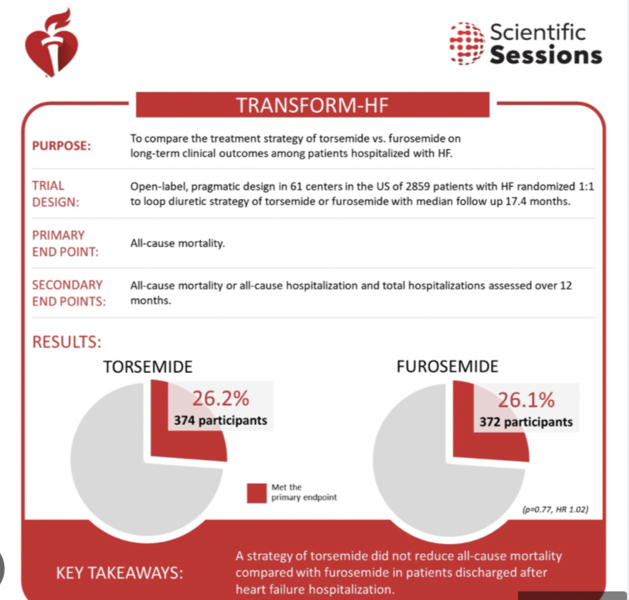
However the TRANSFORM-HF trial presented at the AHA 2022 in Chicago revealed that patients treated for heart failure (HF) with the loop diuretic torsemide had similar rates of death and hospitalization compared with those treated with furosemide, a more commonly prescribed diuretic. This is the first randomised trial comparing the 2 diuretics.


The trial enrolled patients hospitalized with worsening or new-onset HF with a plan for long-term loop diuretic therapy who had either an EF of 40% or lower or, regardless of EF, elevated natriuretic peptide levels when hospitalized.


Overall, 1428 were assigned to receive furosemide as their initial oral diuretic and 1431 patients were assigned to the torsemide-first strategy.


The pragmatic, open label study randomized 2,859 patients at more than 60 U.S. hospitals to either torsemide or furosemide. Of the participants, more than one-third were women, one-third were Black, and the median age was 65 years. Researchers also noted a broad range in the severity of HF ranging from recent diagnosis to worsening stages. Follow-up via phone was conducted at 30 days, six months and 12 months after hospital discharge.


The primary endpoint was all cause mortality, secondary endpoint was all cause mortality or all cause hospitalisation.


After a median of 17.4 months, results showed nearly identical mortality rates in both the torsemide (26.1%) and the furosemide (26.2%) groups. At 12 months, all-cause death and hospitalization rates were also similar in the torsemide (47.3%) and furosemide (49%.3) groups. Researchers noted the primary outcome demonstrating furosemide equivalence to torsemide was the same across all pre-specified subgroups including ejection fraction.


Either furosemide or torsemide are acceptable options for diuresis among hospitalised patients with heart failure, as there is little difference in clinical outcomes over year.


Treating physicians can continue starting such patients with HF on either agent, at their discretion, without concern that the choice may compromise outcomes.


Although furosemide is the most commonly used loop diuretic in HF,  torsemide has both known and theoretical advantages that set it apart. Torsemide is more than twice as potent as furosemide and more bioavailable, and its treatment effect lasts longer.


Torsemide may have pleiotropic effects such as downregulating the renin-angiotensin-aldosterone system (RAAS), reduce myocardial fibrosis and promote reverse ventricular remodeling.


In practice, torsemide may be preferred in patients with furosemide resistance or challenges with bioavailability, especially in very advanced heart failure with gut edema, where oral furosemide is not effectively absorbed.  Torsemide may be a better choice  for individuals who have diuretic resistance with advanced congestion.


Despite theoretical advantage torsemide did not out perform furosemide in acute decompensated heart failure ; it is possible that certain patient groups may respond differently to these 2 diuretics. It is unlikely that there were Indians in the cohorts studied. Both drugs are manufactured in India , ands are quite cheap. 


The researchers concluded that “Among patients discharged after hospitalization for heart failure, torsemide compared with furosemide did not result in a significant difference in all-cause mortality over 12 months. However, interpretation of these findings is limited by loss to follow-up and participant crossover and nonadherence.”


https://jamanetwork.com/journals/jama/article-abstract/2800428

ANTI PLATELET THERAPY AFTER CORONARY STENTING AND INTRACEREBRAL BLEED.


Recently a middle aged diabetic and hypertensive male who had been stented with an everolimus eluting tent in the left anterior descending coronary artery for unstable angina was admitted with acute onset of weakness in his left arm, and systolic blood pressure of 200 mm Hg. The patient was on DAPT consisting of clopidogrel and aspirin (75 mg each). A 128 slice CT scan of brain revealed a right thalamic bleed. The patient was admitted in intensive care, and DAPT stopped. A repeat CT scan did not show any progression in the bleed or cerebral oedema.  There was modest improvement in power of his left arm.This time clopidogrel was resumed minus aspirin, with tight control of blood pressure. The patient was discharged on blood pressure therapy, injection Lantus insulin, and mono anti-platelet therapy ( clopidogrel 75 mg once a day).


The above case illustrates the dilemma faced by a treating cardiologist when a recently stented patient is admitted with a cerebral bleed. Continuing with DAPT risks aggravating the intra cerebral hemmorhage , while stopping anti platelets can result in acute stent thrombosis, which in turn could be lethal. There are no guidelines in management of such patients, apart from a few case reports.


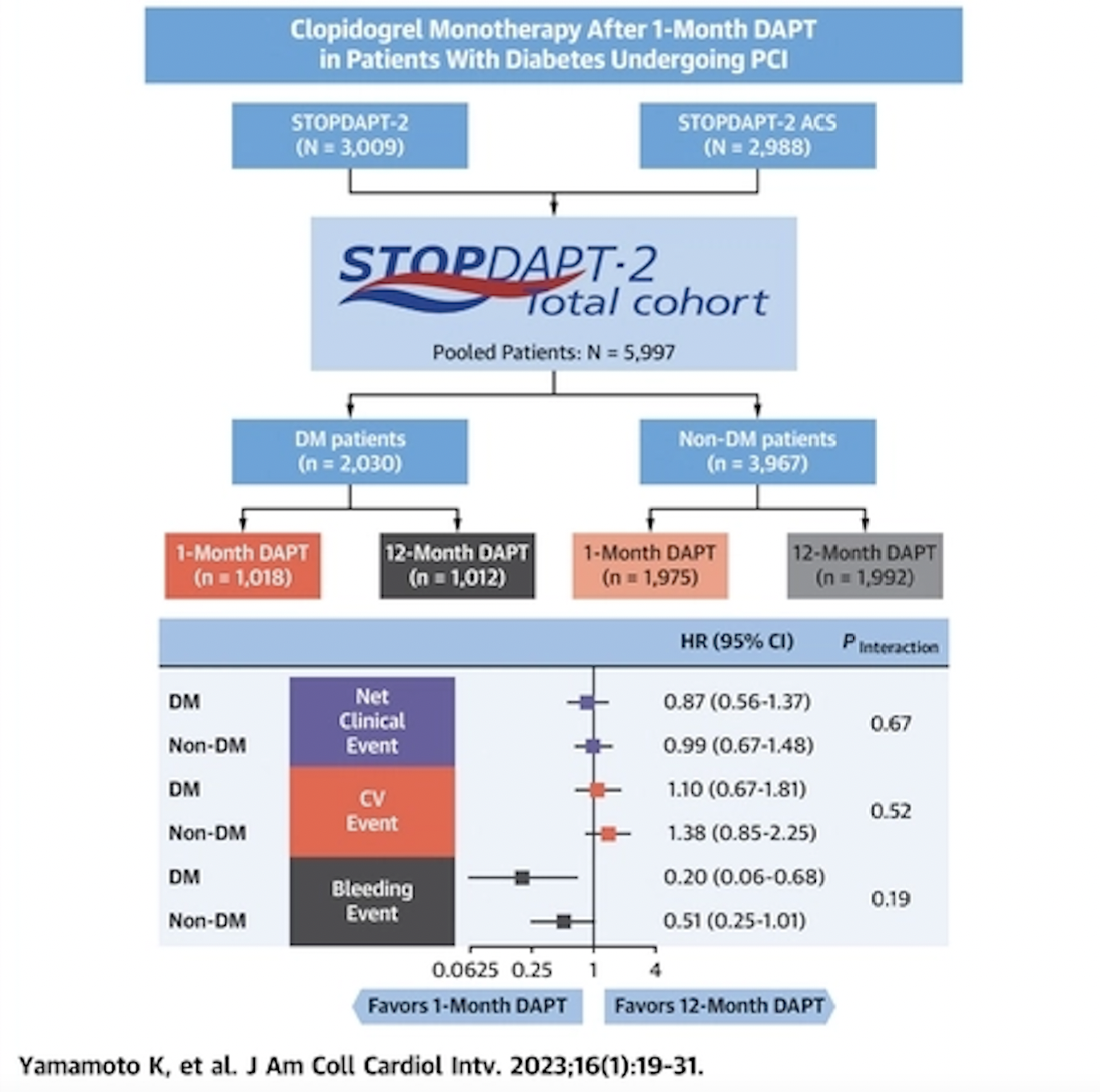
Some light is thrown by a Japanese paper published today in JACC:Cardiovascular Interventions (J Am Coll Cardiol Intv. 2023 Jan, 16 (1) 19–31). The study highlights that clopidogrel mono therapy after just one month of DAPT is safe and effective in patients with diabetes undergoing percutaneous coronary intervention (PCI).


There have been randomised trials showing it is possible to shorten DAPT subsequent to coronary stenting with no difference in cardiovascular outcomes , but significant reduction in bleeding complications. 


The Japanese study is a subgroup analysis of the STOPDAPT-2 trial. The study focused on data from nearly 6,000 PCI patients, including 2,030 patients with diabetes and 3,947 patients without diabetes.


Patients with diabetes were  older and more likely to have a higher BMI. They were also more likely to have a history of prior coronary revascularization, prior myocardial infarction, prior stroke, heart failure, anemia, chronic obstructive pulmonary disease, peripheral artery disease, chronic kidney disease, hypertension or hyperlipidemia.


All patients in the analysis were treated with an initial month of DAPT with aspirin and clopidogrel or prasugrel. Patients were then randomized to continue DAPT for up to 12 months or switch to clopidogrel monotherapy.


The study’s primary endpoint was a composite of cardiovascular events (cardiovascular death, myocardial infarction, definite stent thrombosis or stroke) and bleeding events after one year.


Overall, among patients with diabetes, that endpoint was seen in 3.58% of patients in the clopidogrel monotherapy group and 4.12% of patients in the 12-month DAPT group (p = 0.55) Among patients without diabetes, that endpoint was seen in 2.46% of patients in the clopidogrel monotherapy group and 2.49% of patients in the 12-month DAPT group (p=0.97)


Importantly, the authors noted that there was much less bleeding if the patient was treated with clopidogrel monotherapy (0.30%) as opposed to 12 months of DAPT (1.5%), ( p=0.01).


The limitations were that many eligible patients were not enrolled into the STOPDAPT-2 trial due to “physician choice or patient refusal,” and many of those patients may have faced a higher risk of experiencing a cardiovascular or bleeding event after PCI.


Hence patients in this study represent a somewhat lower risk population than those encountered in real clinical practice. The incidence of the cardiovascular and bleeding events in this study was very low.”


Additional studies are still necessary to determine the optimal monotherapy strategy for diabetic patients undergoing PCI.


The authors conclude “clopidogrel monotherapy after 1-month DAPT compared with 12-month DAPT reduced major bleeding events without an increase in cardiovascular events regardless of diabetes, although the cndings should be considered as hypothesis generating, especially in patients with acute coronary syndrome, because of the inconclusive result in the STOPDAPT-2 ACS trial. (Short and Optimal Duration of Dual Antiplatelet Therapy After Everolimus-Eluting Cobalt-Chromium Stent–2 ).


There is however little data on management of intra-cerebral bleed soon after coronary stenting. Such a patient should be considered a high risk bleed. One option would be to resume mono therapy with clopidogrel as soon as possible provided there is no increase in the bleed. Duration for discontinuation of anti platelet would depend on the individual case, it will have to be shorter in the event of a recent bleed, as chances of stent thrombosis are highest in the 4 to 6 weeks following the index procedure.


Crucially STOPDAPT-2 employed everolimus eluting stents, but GLOBAL LEADERS trial with biolimus eluting stents showed that among patients who underwent PCI with a biolimus-eluting stent, 1 month of DAPT followed by ticagrelor monotherapy for 23 months was noninferior, but not superior to 12 months of DAPT followed by aspirin monotherapy for 12 months. The composite outcome, components of the primary outcome, and major bleeding were similar between treatment groups (Franzone A, McFadden E, Leonardi S, et al. Ticagrelor Alone Versus Dual Antiplatelet Therapy From 1 Month After Drug-Eluting Coronary Stenting. J Am Coll Cardiol 2019;74:2223-34). No difference in bleeding complications could be explained by the greater potency of ticagrelor compared to clopidogrel.


The 3 year followup of the SMART CHOICE trial showed 3-month DAPT,  followed by P2Y12 inhibitor monotherapy was associated with a lower risk of clinically relevant major bleeding than prolonged DAPT. Although the 3-year risk of ischemic cardiovascular events was comparable between the 2 groups, this result should be interpreted with caution owing to the limited number of events and sample size.Bleeding Academic Research Consortium (BARC) types 2–5 bleeding occurred in 3.2% of the shortened DAPT group and in 8.2% of the prolonged DAPT group. Major bleeding, BARC types 3–5, occurred in 1.2% of the shortened DAPT group and in 2.4% of the prolonged DAPT group.


1. Ko Yamamoto, Hirotoshi Watanabe, Takeshi Morimoto, et al. Clopidogrel Monotherapy After 1-Month Dual Antiplatelet Therapy in Patients With Diabetes Undergoing Percutaneous Coronary Intervention. J Am Coll Cardiol Intv. 2023 Jan, 16 (1) 19–31.

ISRAELI PAPER DESCRIBES OMICRON VARIANT INDUCED MYOCARDITIS DESPITE BEING TRIPLE mRNA JABBED.


https://academic.oup.com/ehjcr/article/6/10/ytac407/6748255


Israeli paper published in European Heart Journal (October 2022) reports 2 cases of myocarditis following infection with Omicron variant.


One case lands up in hospital with pule less ventricular tachycardia at karate of 250/min In both cases PCR and sequencing confirmed infection by Omicron variant.


However , both patients had been triple jabbed with Pfizer vaccine ( last injection given in August 2021).


Are the researchers of this paper implying that people need yet another (fourth) Pfizer vaccine jab to prevent infection ?


Or is it their message that despite 3 vaccine injections you can still get infected by SARS CoV 2 and need hospitalisation?


DAMAR HAMLIN COLLAPSED DUE TO COMMOTIO CORDIS OR CONTUSIO CORDIS; NOT NECESSARILY BECAUSE OF VACCINE INJURY.


https://www.ahajournals.org/doi/full/10.1161/circep.111.962712


Undoubtedly excess all cause mortality is occurring in mRNA vaccinated countries; there has been a significant increase in deaths since the jabs began. Omicron or its sub variants do not explain the surge in deaths as they are less virulent albeit more infective. The needle of suspicion points to vaccines.


However, Damar Hamlin’s collapse is best explained by the phenomenon of Commotio Cordis , where physical impact to the left chest can spark lethal ventricular arrhythmia.


In the cricket “Body Line ” series in the 1930’s the Aussie captain suffered a blow to his chest, collapsed, and luckily for all concerned recovered on this own.


Most importantly, let us pray Damar recovers fully.

GERMAN AUTOPSY STUDY CONFIRMS VACCINE INDUCED MYOCARDITIS

A German autopsy study has found 5 patients ( of 25 sudden deaths soon after covid vaccination ) with sudden death within 3 days of mRNA covid vaccination demonstrating myocarditis or inflammation of heart. Mean age of those dying was 58 years. These 5 dead autopsied hearts did not have evidence of any viral infection, or underlying heart disease ( coronary artery disease or cardiomyopathy).


A few months ago JAMA published official American (CDC) showing an incidence of 105 per million cases of myocarditis post Covid vaccination. Mean age is 21 years, 80 % are males, and onset of myocarditis was 2 days post injection of vaccine. Almost 98% had raised troponin levels, more than 70% had ECG and cardiac MRI changes.


Last year a scientific letter published in the New England Journal of Medicine described 2 patients with severe left ventricle dysfunction soon after mRNA jabs, both were in their 40’s. The left ventricle ejection fraction dropped to 15-20% only, there was no evidence of any viral infection, their coronary arteries were normal. One of them went into cariogenic shock and died.


The German autopsy researchers suggest that post vaccine myocarditis may be due to an auto immune reaction as there was a preponderance of CD4 type T cells apart from heart cell damage. They postulate that because heart size was normal , the sudden deaths may have been to rhythm disturbance, either the heart stopped beating or there was lethal ventricular tachycardia/ventricular fibrillation.


Moreover inflammation in these 5 sudden death hearts ( in the German study) was restricted to the right ventricle or inter ventricular septum.


1) Autopsy‐based histopathological characterization of myocarditis after anti‐SARS‐CoV‐2‐vaccination. Clinical Research in Cardiology https://doi.org/10.1007/s00392-022-02129-5 https://link.springer.com/article/10.1007/s00392-022-02129-5


2) Myocarditis after Covid-19 mRNA Vaccination n engl j med 385;14 nejm.org September 30, 2021 https://www.nejm.org/doi/full/10.1056/NEJMc2109975


3)Myocarditis Cases Reported After mRNA-Based COVID-19 Vaccination in the US From December 2020 to August 2021 JAMA. 2022;327(4):331-340. doi:10.1001/jama.2021.24110 https://jamanetwork.com/journals/jama/fullarticle/2788346


EXAMINING THE INDIAN CRICKET TEAM OR EXCESS DEATHS IS BLASPHEMY


The performance of the Indian cricket team in the T20 semifinal was a downright disgrace but no cricket pundit Indian or foreign dare utter a word for fear of swift retribution by the BCCI. No more TV appearances, no more commentary, no more newspaper column, no more perks.


Despite having the fastest bowler in the world to venture into Australian pitches for the T 20 World Cup beggars belief, an astounding decision/blunder by any standards.


We had the worst Powerplay average score (runs scored in the first 6 overs)! So much for our much acclaimed opening pair. Whatever happened to Prithvi Shaw, Gill, Ishan Kishan, and of course Sanju Samson ??


The worlds 100 m (men) record holder Usain Bolt despite being a Jamaican considered Waqar Younis his favourite fast bowler ! Waqar Younis, one of the greatest fast bowlers the world has ever seen, was picked up for the Pakistani international team by Imran Khan as he watched him play a first class match on television! Waqar had played a mere 6-7 first class matches by then. The BCCI must make note of the above.


Similarly no one seems to be notice excess non Covid deaths in many parts of the world, especially where gene vaccines have been employed. In England and Wales excess deaths have occurred week after week since April 2022, there are now more than 39,000 excess deaths. Less than 6% of excess deaths are Covid related. The UK government statistics bureau (ONS) provides data ( on their website) on deaths by vaccine status; regardless of age more people who are vaccinated have died than those unvaccinated.


It is imperative that the causes of excess deaths across the planet are ascertained before mandating boosters on a yearly or 6 monthly basis. Whether these excess deaths are collateral damage because of lockdowns, or due to vaccines needs to found out, and quickly. There are too many elephants in the room.

EMPAGLIFLOZIN FOR DIABETES , HEART FAILURE , AND KIDNEY FAILURE.


The SGLT2 inhibitor Empagliflozin is well documented to significantly reduce cardiovascular death and hospitalisation for heart failure when compared to placebo, in patients with diabetes having high risks for heart disease.


Empagliflozin also significantly reduced hospital admission or cardiovascular mortality in patients with heart failure with both reduced or preserved ejection fraction; in HFrEF and HFpEF. The EMPEROR REDUCED and EMPEROR PRESERVED trials that included more than 9000 patients with heart failure and randomised them to 10 mg Empagliflozin or placebo and demonstrated substantial effectiveness in cutting hospital admission for heart failure or heart disease death.


Three days ago a large double blind randomised trial EMPA KIDNEY including 6600 patients with chronic kidney disease showed significant slowing of kidney disease (by 28%), with 10 mg of Empagliflozin once. day. The drug was effective in patients regardless of their sex, ethnicity or if they had diabetes or not. Empagliflozin therapy led to a lower risk of progression of kidney disease or death from heart disease than placebo.


There was also a 14% reduction in hospitalisation for heart failure.


Empagliflozin is available in India, the dose is 10 mg tablet once a day, and the cost is quite reasonable.

DEATH DURING SLEEP IS NOT ALWAYS DUE TO VACCINE INDUCED MYOCARDITIS


The CDC chief stated last month that it is “critically important” one takes the bivalent vaccine injection, but in a month she came down with Covid.


A senior Pfizer executive has gone on record in the European Parliament that Pfizer was completely in the dark of their vaccine stopping transmission before putting the vaccine in the market; Pfizer had absolutely no data on transmission ! Considerable data has been collected demonstrating adverse effects of mRNA and DNA vaccines in young people, who any way are naturally protected from severe covid, hence vaccinating the young is Illogical.


Hence, quite a few countries, are now not keen on vaccinating every one under the age of 50 (Denmark) or even 65 (Norway). Myocarditis following mRNA vaccines is a real and present danger, with inflammation persisting for months as shown by cardiac MRI.


There are also reports of people dying in their sleep.An article in the Epoch Times (a far right anti communist news paper) has quoted a senior American cardiologist that deaths during slumber are because of vaccine myocarditis. There is a “paradoxical” release of catecholamines that triggers lethal arrhythmia resulting in death.


I am sceptical of this explanation, that all deaths during sleep are due to myocarditis subsequent to mRNA vaccination.


It is imperative that Long QT Syndrome type 3 or LQT3 is ruled out in deaths whilst asleep. LQT3 is a well recognised genetic disease with gain of function in the gene encoding sodium channel.


In this channelopathy there are mutations in the SCN5A gene present in Chromosome 3. People afflicted with this genetic disease can die while asleep or at rest, there is no role of emotional or physical stress.


Long QT syndromes has prolongation in the recharging or depolarisation time of the heart. When the QT interval on the 12 lead ECG gets longer than 500 ms the concerned  person is vulnerable to lethal polymorphic ventricular tachycardia that swings around a point and therefore called “Torsades de Pointes” or TdP.


Patients with LQT1 and LQT2 suffer from palpitations, fainting spells, seizures, and sudden death. Physical effort such a swimming or diving into a pool can trigger TdP in LQT1, while a sudden loud noise does it in LQT2.


But LQT3 albeit uncommon can spark a lethal polymorphic ventricular tachycardia during sleep. Patients with LQT usually but not always have a prolonged QT interval in the ECG, and the problem is confirmed by genetic analysis, which is expensive and takes time.


In conclusion before damning vaccine induced myocarditis for every death during sleep, LQT3 should and must be considered. The molecular analysis can be done at autopsy.


Especially because a sibling or a relative could be the next casualty.


PS: The Epoch Times has some excellent reporting on Covid 19, and I highly recommend it.

DO NOT USE PFIZER’S PAXLOVID IF YOU HAVE HEART DISEASE


The NIH calls Paxlovid “the only highly effective oral anti viral” treatment available in its Covid treatment guidelines.


However, the NIH concedes Paxlovid ( a combination of ritonivir and nirmatrelvir) is not of any benefit in standard risk patients with Covid 19.


Also, Paxlovid , is associated with a “rebound ” effect in some patients despite apparent clearance of clinical symptoms. Tony Fauci ,four times jabbed , managed to still get infected with Covid 19. Fauci, therefore took his much touted Paxlovid, but landed up with a rebound infection, that was more severe than the previous one ( according to Fauci himself).


The third problem with Paxlovid is that it has serious interactions with other drugs; unfortunately just about every drug used in heart medicine has to be either stopped completely or dose adjusted. The list of drugs affected with Paxlovid includes antiplatelet drugs such as clopidogrel and ticagrelor, anti coagulants, statins, anti arrhythmic, anti hypertension medicines, and also heart failure medicines. There may be serious adverse effects if these drugs are not withheld or dose adjusted. Please check the NIH guidelines on Covid 19 treatment that is freely available on the internet.


I as a cardiologist would therefore not advise Paxlovid to my Covid 19 patients.


“Although ritonavir-boosted nirmatrelvir demonstrated a clinical benefit during the EPIC-HR trial, the benefits in unvaccinated patients who are at low risk of progression to severe disease or in vaccinated people who are at high risk for progression to severe disease are unclear. The EPIC-SR trial, which included both of these populations, found that ritonavir-boosted nirmatrelvir did not reduce the duration of symptoms and did not have a statistically significant effect on the risk of hospitalization or death compared to placebo, although the event rates were low.” NIH Covid 19 treatment guidelines ;page 191.


Pfizer is looking at a $ 22 billions annual business with Paxlovid.

IS 3 MONTHS OF DAPT AFTER STENTING ENOUGH ? SMART CHOICE SAYS YES.


Andreas Gruntzig performed the first PTCA on 16th September 1977, this was a 38 years old smoker who was alive and well till 2018, in the meantime he had a bare metal stent stent inserted in 2000 and then a drug eluting stent in 2014, he had had a left anterior descending coronary artery block to begin with.


The second PTCA was done also by Gruntzig on 18th October 1977 in a 44 year old male, who had blocks in his left main and right coronary arteries. The procedure this time was not successful and the patient needed a CABG operation in 1978, but all his grafts got blocked by 1997 and so he had a repeat CABG operation.


Following a drug eluting stent implantation it becomes necessary to begin blood thinners called antiplatelets. Two antiplatelets have  used conventionally, that is a combination of aspirin and a P2Y12 inhibitor ( clopidogrel, ticagrelor, or prasugrel). This strategy is also called dual anti platelet therapy or DAPT. DAPT prevents stent thrombosis that can be catastrophic and fatal. Till recently DAPT was administered for at leat a year and then the P2Y12  inhibitor was stopped, but aspirin was continued for life. The problem was that DAPT or aspirin alone could serious bleeding complications. 


Recently randomised trials comparing DAPT for 1-3 months versus DAPT for one year have shown that risks for death, heart attack or stroke are the same, but bleeding complications are significantly less with shortened DAPT duration.


A meta analysis published last year in the BMJ that included 6 randomised trials with almost 24,000 patients concluded the same, shorter DAPT was as good as one year of DAPT, with substantially less bleeding complications. It was also seen that women were especially at an advantage with the short DAPT, because they had significantly less mortality.


Now another randomised study from Korea in 3000 patients who were stented with second generation drug eluting stents (published late September 2022 in JAMA Cardiology) has shown that 3 months of DAPT  has similar clinical risks as longer DAPT. Crucially, this study SMART CHOICE has the longest follow up of 3 years.


SMART CHOICE concludes that 3 months of DAPT is as good as 3 years of DAPT regarding death, myocardial infarction and stroke , but with almost 50% less severe bleeding complications. In this study as also in similar randomised rials the shorter DAPT regimen continues with a  P2Y12 blocker while aspirin is dropped; this cohort is compared with 3 years of continuous DAPT.


The take home message is that 3 months of DAPT after a second generation drug eluting stent is enough, with continuation of clopidogrel, ticagrelor or prasugrel, but dropping aspirin.

TRY ENTRESTO AND AN SGLT2 INHIBITOR FOR 6 MONTHS BEFORE A MITRAL EDGE TO EDGE PROCEDURE (TEER)


Mitral regurgitation accompanying heart failure is a bad problem. Quality of life goes down, hospitalisation goes up, and life gets shortened. In HFrEF the left ventricle dilates and with this the papillary muscles get displaced and the 2 mitral valve leaflets cannot coat or close adequately. Net result is blood gets thrown back with each contraction of the heart and heart failure worsens. This mitral regurgitation is defined as secondary or functional MR.


Unfortunately surgical repair or surgical valve replacement has not been very successful. A device called trans catheter end to end repair has been around for almost a decade and at least one randomised trial done in the US has shown excellent results , in fact some have called them spectacular. 600 patients with moderate and severe MR were randomised to the TEER procedure or to guideline directed medical therapy. At 2 years mortality had been reduced by 37% and hospitalisation by 47% in the TEER group.


With TEER a catheter is negotiated from a groin vein into the right atrium, and from there into the left atrium by puncturing the inter atrial septum, and finally the nitrate leaflets are clipped. The clipping reduces or removes mitral regurgitation. 


But unlike the American study a French (also randomised) failed to show any difference in events with TEER compared to guideline directed medical therapy. American researchers explained lack of efficacy because of less sick patients in the French trial. 


A sub analysis of the PROVE-HF trial was published in Circulation a week ago, that shows the number of patients with moderate or severe mitral regurgitation halved at 6 months when they were given sacubitril/valsaratan treatment. The salutatory results persisted for 12 months. It must be noted that earlier PROVE-HF had reported an absolute increase of 9.5% in left ventricle ejection fraction on an average at 12 months on sacubitril/valsartan therapy. 


Crucially the American (COAPT) trial on TEER had less than 3% patients on sacubitril/valsartan while the French trial had 12.1% on the combination medicine. The patients were obviously not on optimal guideline directed medical therapy.


Hence, in patients with severe heart failure (HFrEF) accompanied by severe mitral regurgitation a trial of drug therapy with sacubitril/valsartan should be tried, because half of these patients may respond and not need the TEER device that is expensive and not without some morbidity. In case the patient does not respond to drug therapy, TEER should be suggested to the patient. It would be a good idea to add an SGLT2 inhibitor to sacubitril/valsartan while waiting for 6 months.


Also an US observation registry has shown a 50% reduction in mortality with TEER in patients with cariogenic shock accompanied by mitral regurgitation at one year follow up. This is not a randomised study but s the observations are hypothesis generating apart from being reassuring. So far nothing has really worked in cariogenic shock, which has a mortality of 50% at 6 months.

THE GRADE TRIAL ON LOW RISK PATIENTS WITH DIABETES


Today October 4 is the second birthday of my dear granddaughter Anushka, so a very Happy Birthday to Anushka Baba.


India has a huge burden of patients with diabetes, there were about 77 million in 2019. The highest prevalence state wise was 13.6% in Chandigarh, with Tamil Nadu having 10.4% and Punjab 10%. Delhi had a prevalence of 26% and Chennai 22%.


Metformin is unanimously considered the first choice in treating type 2 diabetes, and recent randomised cardiovascular outcome trials have shown effectiveness of SGLT2 inhibitors and GLP receptor agonists in improving clinical outcomes. These studies were done in patients with established cardiovascular disease or risk factors.


Treatment of low risk diabetes patients however remains unclear. The GRADE trial attempts to fill the gaps. The trail included more than 4000 patients already on metformin, who had onset of diabetes in the last 10 years. Four classes of drugs were added to metformin. Insulin glargine (long acting), a sulfonylurea namely glimepiride, a GLPR agonist , or a DPP4 inhibitor namely sitagliptin. At base line glycated haemoglobin ranged from 6.8 to 8.5%. The aim in treatment of diabetes is achieving a glycated haemoglobin below 7%.


At an average follow up of 5 years adding insulin glargine to a GLP R agonist was more effective in reducing glycated haemoglobin. Sitagliptin was the least effective in reducing glycated haemoglobin. Hypoglycemia was most at 2.2% with glimepiride. Microvasular complications ( retinopathy, nephropathy, neuropathy ) were similar, but liraglutide reduced cardiovascular outcomes by 29%.


A limitation is that an SGLT2 inhibitor was not included because this class of drug was not approved by the FDA when the GRADE trial began.


The take home message is that in a low risk patient with diabetes begin treatment with metformin, and then add an SGLT2 inhibitor or a GLPR agonist or if need be a long acting insulin.


Importantly a sulfonylurea like glimepiride would also be effective in controlling glycated haemoglobin level. 


All these drugs are available in India at a very reasonable price.


But diabetes is a progressive disease as the GRADE trial has shown with almost 70% pf patients having a glycated haemoglobin above 7% at 5 years.

DO NOT FORGET THE SACUBITRIL VALSARTAN COMBO WHEN TREATING HEART FAILURE


It is more or less clear that an SGLT2 inhibitor will significantly reduce hospitalisation or cardiovascular mortality in patients both with reduced or preserved ejection fraction. We have the Emperor Reduced, Emperor Preserved, Data HF, and Deliver Trials to confirm this. We also know that there are lakhs of patients with heart failure, with half of them having  preserved ejection fraction. The Deliver Trial reported efficacy of dapapgliflozin in ejection reactions more or less than 60%, the DAPA heart failure trial had shown efficacy in an ejection fraction less than 40%. But there is one important point very few patients were on ARNI class of medicine in the SGLT2 trials, just around 15%. I will come to ARNI very soon.


Sadly heart failure is a progressive disease with 50% patients needing re-hospitalisation in a month and 20% dying in 2 years. This is despite therapy. We have a huge problem on our hands. Over the years multiple randomised trials showed that certain drugs significantly reduce mortality in heart failure patients. Beta blockers reduced death by 30%, a mineralocorticoid receptor blocker by 30%, and an ACE inhibitor by 25%.


Against this background the PARADIGM heart failure trial presented in 2014 was a landmark study that included more than 8400 patients with heart failure and median ejection fraction of only 29%. Around 22% were in NYHA class II, 80% were men, average age was 67 years. Follow up was for 27 months. The study randomised patients to an ARNI or an ACE inhibitor called enalapril. An ARNI is an angiotensin receptor neprilysin inhibitor with sacubitril and valsartan. Sacubitril is the neprilysin blocker and valsartan the AT receptor blocker.


By 27 months the ARNI treated group had a significant 20% reduction in cardiovascular death or hospitalisation for heart failure as compared with the enalapril group. This was a robust difference as these patients were already on a beta blocker, a diuretic, an MRA and 7% had a biventricular pace-maker.


Another trial called PROVE followed 800 patients with an ARNI for one year. There was a significant fall in NTproBNP levels that started falling as early as 2 weeks from treatment onset, and crucially the ejection fraction increased on an average by 9.5% at one year. Such a rise in ejection fraction compares with that of a biventricular pacemaker treatment.


So in conclusion a chronic heart failure patient should be ideally treated with an ARNI, a beta blocker, an MRA and an SGLT2 inhibitor.

VERICIGUAT FOR WORSENING HEART FAILURE


Current heart failure guidelines recommend using 4 heart failure drugs simultaneously in patients with chronic heart failure and reduced ejection fraction to get the best results. There could be in fact a 74% reduction in mortality if all 4 drugs are employed; these are a beta blocker, an ACEI or ARNI, an MRA and an SGLT2inhibitor.


The American guidelines mention briefly that in the event of worsening heart failure despite guideline mandated therapy a new drug named vericiguat be used as the Victoria Trial has reported a significant 10% reduction in the primary end point of cardiovascular mortality or heart failure admission in the cohort randomised to vericiguat as compared to placebo.


The Victoria Trial studied 5050 patients of chronic heart failure with educed ejection fraction who worsened in the previous 6 months ( needed hospital admission or injection of a diuretic). The median ejection fraction was 29%, there were 76% males, almost 90% were on 2 guideline medication, while 60% were on triple heart failure drugs. There was modest clinical improvement in a flow up of slightly more than 10 months, with he curve diverging at 3 months.


The dose used was 2.5 mg orally once a day to be titrated to 10 mg once a day.


One tablet of 2.,5 mg vericiguat costs about Rupees 100 in India.


A sub analyses presented in the ACC Meeting this year highlighted that vericiguat was most effective in the cohort of patients with an LV ejection fraction below 24%.


There were more cases of symptomatic hypotension, syncope and anaemia in the vericiguat group but kidney function and electrolytes were unaffected.


I would use the the 4 heart failure medicines mentioned above , before administering vericuguat in a patient with heart failure and reduced ejection fraction (HFrEF).


Also, it cannot be emphasised that despite the American Heart Association recommending vericiguat in worsening heart failure , there was no significant reduction in mortality in the treated group, albeit follow up was less than 11 months.